Two-hundred and ninety-five healthy individuals (51.6% male) between the ages of 8.1 and 68.1 years (mean 29.72 ± 14.31; median 25.97). All subjects received a DTI exam at the North Shore University Medical Center, Manhasset, New York, on a GE Signa HDx 3.0 T system (General Electric, Milwaukee, Wisconsin). The sequence included volumes with diffusion gradients applied along 31 nonparallel directions (bvalue: 1000 s/mm2) and five volumes without diffusion weighting (repetition time: 14 seconds, echo time = minimum, matrix : 128 x 128, field of view: 240 mm). Each volume consisted of 51 contiguous 2.5-mm axial slices acquired parallel to the anterior-posterior commissural line using a ramp sampled, double spin-echo, single shot echo-planar imaging method. All scans were reviewed by a radiologist, and all images were visually inspected to ensure that no gross abnormalities or artifacts were evident. Image processing was conducted using the Functional Magnetic Resonance Imaging of the Brain Software Library (FSL version 5.1; Oxford, United Kingdom; http://fsl.fmrib.ox.ac.uk/fsl). Eddy-current induced distortions and head-motion displacements were corrected through affine registration of the 31 diffusion volumes to the first b0 volume using FSL’s Linear Registration Tool. The b-vector table (i.e., gradient directions) for each participant was then adjusted according to the rotation parameters of this linear correction. Non-brain tissue was removed using FSL’s Brain Extraction Tool
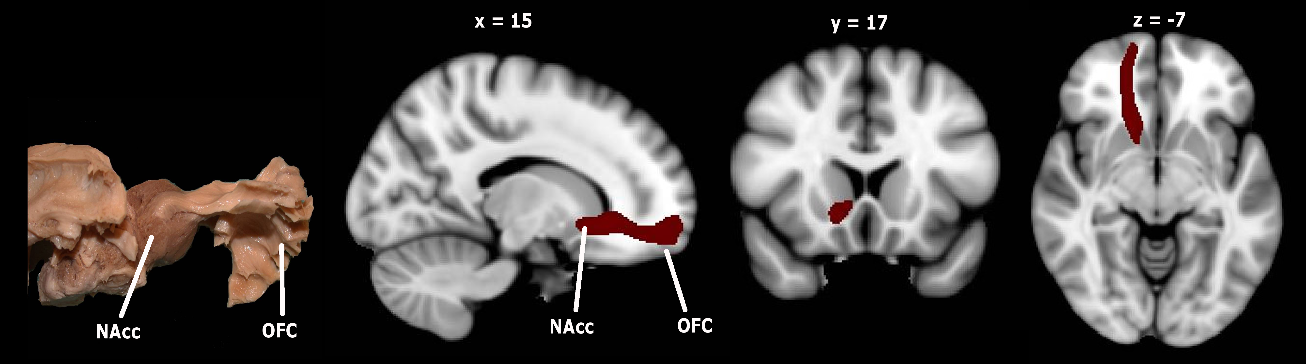
The probable trajectory of the accumbofrontal tract was traced as follows. Within-voxel probability density functions of the principal diffusion direction were estimated using Markov Chain Monte Carlo sampling in FSL’s BEDPOSTX tool (Behrens et al., 2003) A spatial probability density function was then estimated across voxels based on these local probability density functions using FSL’s PROBTRACKX tool (Behrens et al., 2003), in which 5000 samples were taken for each input voxel with a .2 curvature threshold, .5-mm step length, and 2000 steps per sample. For each tract, seed masks, waypoints, termination and exclusion masks were defined on the MNI152 T1 1-mm template. For the accumbofrontal tract, exclusion maps included the entire contralateral hemisphere, superior frontal regions, and regions posterior to the striatum, and the seed masks were Harvard-Oxford atlas defined ROIs of the NAcc and OFC. Masks were normalized to each subjects’ diffusion space using FSL’s Linear Registration Tool (Jenkinson and Smith, 2001) applying the affine parameters obtained by coregistering the first b0 volume to the MNI152 T1 1-mm template. The resulting tracts were thresholded at a normalized probability value of .01 and visually inspected to confirm successful tracing in each individual subject. Mean FA of the entire tract was then extracted for analysis. The fronto-parietal connection, the SLF, was also traced, as a comparison tract that is involved in executive functioning, and the methods for SLF extraction are described in Peters et al (2014).